Back Muscles Anatomy Ct : Back Pains Midback Pains Neck Pains Max Superspecialty Ortho Clinic - Able to move the upper limb as they originate at the vertebral column and insert onto either the clavicle, scapula or humerus.
byAdmin•
0
Back Muscles Anatomy Ct : Back Pains Midback Pains Neck Pains Max Superspecialty Ortho Clinic - Able to move the upper limb as they originate at the vertebral column and insert onto either the clavicle, scapula or humerus.. Includes latissimus dorsi, the trapezius, levator scapulae and the rhomboids. This mri chest (thorax) axial cross sectional anatomy tool is absolutely free to use. Page 156:abdominal wall muscles normal anatomy, axial ct 6 of 9lineslines and labels. (2017, elsevier) should be consulted. Intermediate layer of back muscles.
Muscles of the lumbar spine. The suboccipital muscles (splenius muscle, semispinalis muscles of the neck and head and interspinous neck muscles.) the muscles of the back with the surface (trapezius, latissimus dorsi, thoracolumbar fascia, deltoid) and intermediate layers (serrated muscles, external and internal oblique muscle). Filed under anatomy, ct, head and neck. Radiographers suggest an abdominal ct scan to look for the following: Muscles that attach from the pelvis to the trunk and cross the lumbosacral joint muscles that attach from the pelvis to the thigh/leg and cross the hip joint pelvic floor muscles that are located wholly within the pelvis
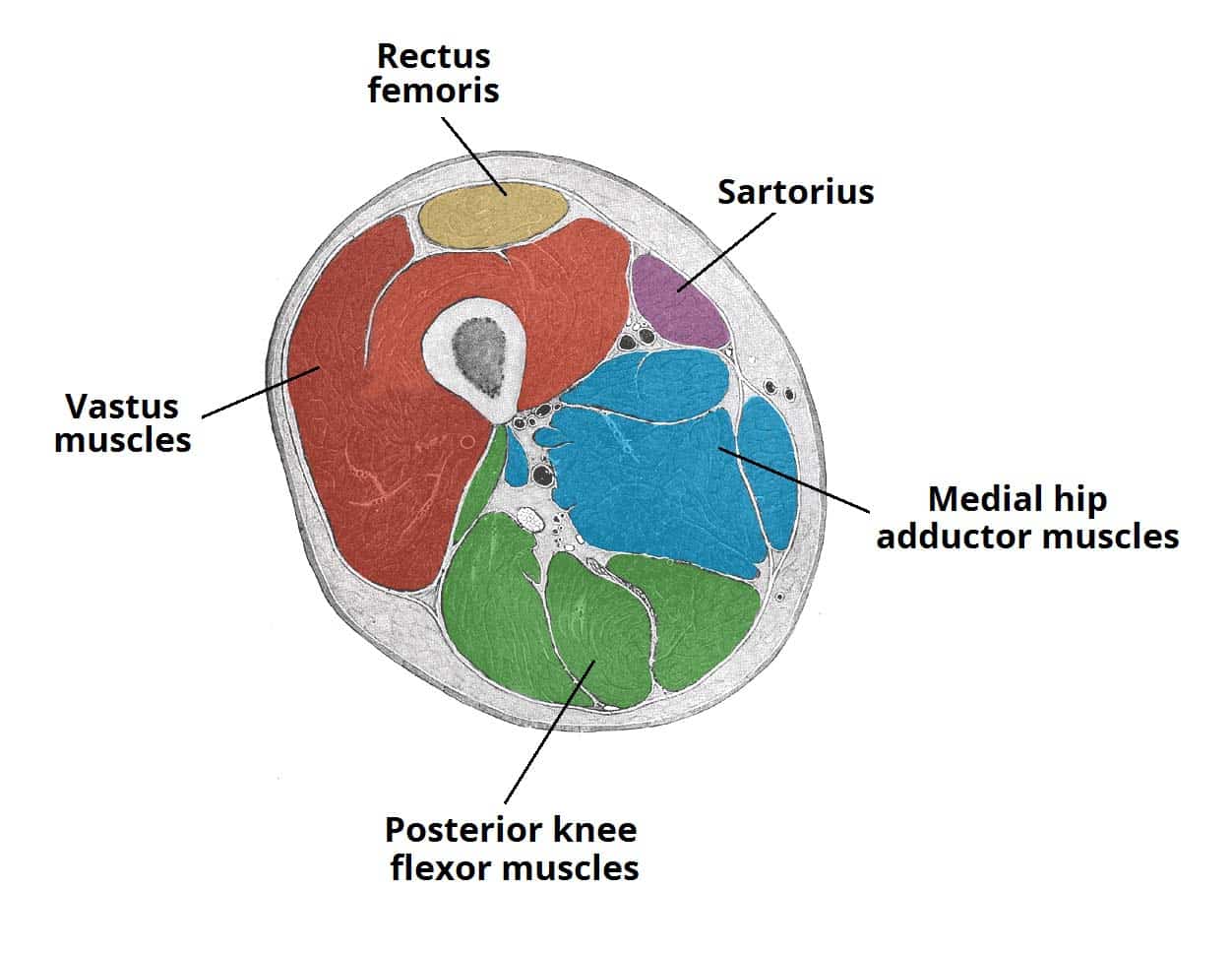
Muscles Of The Anterior Thigh Quadriceps Teachmeanatomy from teachmeanatomy.info Back muscles the muscles of the back are a group of strong, paired muscles that lie on the posterior aspect of the trunk. These important muscles control many motions that involve moving the arms and head — such as throwing a ball, looking up at the sky, and raising your hand. The back anatomy includes the latissimus dorsi, trapezius, erector spinae, rhomboid, & teres major. All these muscles are therefore associated with movements of the upper limb. Posted by radiologypics ⋅ march 21, 2013 ⋅ 1 comment. The seventh cervical vertebra, referred to as c7, meets the first of 12 thoracic vertebrae t1 at the base of the neck, a. The muscles of the back can be arranged into 3 categories based on their location: Superficial muscles of the back want to learn more about it.
Identify the movement and function of the intrinsic skeletal muscles of the back and neck, and the skeletal muscles of the abdominal wall and thorax.
Each block is separated by a disc that sits in between and each vertebra has a facet joint on either side. To perform clinical clinical orthopedic manual therapy to the lumbar spine. The muscles of the back can be arranged into 3 categories based on their location: Related posts of muscles of the lower back and hip diagram muscle anatomy posterior. The extrinsic back muscles are located in the back, but act to produce movements of the shoulder and assist respiration. Iliocostalis subgroup is the most lateral longissimus subgroup is between iliocostalis and spinalis Lumbar aponeurosis and vertebral fascia).—the lumbodorsal fascia is a deep investing membrane which covers the deep muscles of the back of the trunk. They provide movements of the spine , stability to the trunk, as well as the coordination between the movements of the limbs and trunk. 10 rectus abdominus muscle this muscle is consider. Along it are easily palpable spinous processes by palpation of the cervical vii and all lying. The erector spinae group is the intermediate layer of the intrinsic muscles of the back. The neck consists of seven cervical vertebrae, the building blocks of the spine. The seventh cervical vertebra, referred to as c7, meets the first of 12 thoracic vertebrae t1 at the base of the neck, a.
The erector spinae group is the intermediate layer of the intrinsic muscles of the back. This group is made of three subgroups, with the group divisions occurring by location. Includes latissimus dorsi, the trapezius, levator scapulae and the rhomboids. The neck consists of seven cervical vertebrae, the building blocks of the spine. Muscles of the lumbar spine.
Radiological Anatomy X Ray Ct Mri Kenhub from thumbor.kenhub.com Larynx anatomy ct and mri. • its lower end is at the lower border of the cricoid cartilage. Superficial muscles of the back want to learn more about it. Muscles of the lumbar spine. Identify the movement and function of the intrinsic skeletal muscles of the back and neck, and the skeletal muscles of the abdominal wall and thorax. Lower back pain is a pervasive symptom. This blog post article is an overview of the muscles of the lumbar spine of the trunk. Abdominal computed tomography (ct) is a type of medical imaging procedure used to diagnose and monitor internal stomach issues, like cancer, bowel obstruction, and abdominal pain.
On this page youll learn about each of these muscles their locations and functional anatomy.
The back muscles are divided into two large groups: The back is subdivided into the upper, middle, and lower back. The muscles of the chest and upper back occupy the thoracic region of the body inferior to the neck and superior to the abdominal region and include the muscles of the shoulders. The seventh cervical vertebra, referred to as c7, meets the first of 12 thoracic vertebrae t1 at the base of the neck, a. The trapezius muscle is a large, broad superficial muscle of the posterior neck and back. Identify the movement and function of the intrinsic skeletal muscles of the back and neck, and the skeletal muscles of the abdominal wall and thorax. Superficial muscles of the back want to learn more about it. Lumbar aponeurosis and vertebral fascia).—the lumbodorsal fascia is a deep investing membrane which covers the deep muscles of the back of the trunk. Able to move the upper limb as they originate at the vertebral column and insert onto either the clavicle, scapula or humerus. Unknown case 25 hand radiograph. Learn about the superficial, intermediate and deep muscles of the back. Page 156:abdominal wall muscles normal anatomy, axial ct 6 of 9lineslines and labels. Sign up for your free kenhub account today and join over 1078234 successful anatomy students.
The superficial back muscles are covered by skin, subcutaneous connective tissue and a layer of fat. Page 156:abdominal wall muscles normal anatomy, axial ct 6 of 9lineslines and labels. On this page youll learn about each of these muscles their locations and functional anatomy. Superficial back muscles, intermediate back muscles and intrinsic back muscles.the intrinsic muscles are named as such because their embryological development begins in the back, oppose to the superficial and intermediate back muscles which develop elsewhere and are therefore classed as extrinsic muscles. The muscles of the chest and upper back occupy the thoracic region of the body inferior to the neck and superior to the abdominal region and include the muscles of the shoulders.
Http Pdf Posterng Netkey At Download Index Php Module Get Pdf By Id Poster Id 119484 from Superficial muscles of the back want to learn more about it. The muscles of the back can be arranged into 3 categories based on their location: Abdominal computed tomography (ct) is a type of medical imaging procedure used to diagnose and monitor internal stomach issues, like cancer, bowel obstruction, and abdominal pain. The neck consists of seven cervical vertebrae, the building blocks of the spine. The erector spinae group is the intermediate layer of the intrinsic muscles of the back. Larynx anatomy ct and mri. Intermediate layer of back muscles. To perform clinical clinical orthopedic manual therapy to the lumbar spine.
The muscles of the chest and upper back occupy the thoracic region of the body inferior to the neck and superior to the abdominal region and include the muscles of the shoulders.
Identify the movement and function of the intrinsic skeletal muscles of the back and neck, and the skeletal muscles of the abdominal wall and thorax. It gains its name from its diamond shape. The back is subdivided into the upper, middle, and lower back. Unknown case 25 hand radiograph. Includes latissimus dorsi, the trapezius, levator scapulae and the rhomboids. Page 156:abdominal wall muscles normal anatomy, axial ct 6 of 9lineslines and labels. The suboccipital muscles (splenius muscle, semispinalis muscles of the neck and head and interspinous neck muscles.) the muscles of the back with the surface (trapezius, latissimus dorsi, thoracolumbar fascia, deltoid) and intermediate layers (serrated muscles, external and internal oblique muscle). Along with sternocleidomastoid muscle, it is invested by the superficial layer of the deep cervical fascia, which splits around it. Abdominal computed tomography (ct) is a type of medical imaging procedure used to diagnose and monitor internal stomach issues, like cancer, bowel obstruction, and abdominal pain. The back muscles are divided into two large groups: Iliocostalis subgroup is the most lateral longissimus subgroup is between iliocostalis and spinalis The trapezius muscle is a large, broad superficial muscle of the posterior neck and back. It is a complex job to balance the body on two feet and walk upright.
Anatomy of the upper back muscles back muscles anatomy. Along with sternocleidomastoid muscle, it is invested by the superficial layer of the deep cervical fascia, which splits around it.